How to Choose 4D vs 3D Ultrasound for Pregnancy 2026?

Time:2026-10-12 Author:Oliver
0%

Choosing between 3D and 4D ultrasound in 2026 requires more than comparing attractive images. Both technologies create three-dimensional views of fetal anatomy. 4D adds real-time movement, showing a baby turning, stretching, or opening its mouth. That difference can feel remarkable in a dim examination room. Yet, it does not automatically mean better medical care.

So, why is 4d ultrasound better than 3d for pregnancy? In selected cases, live imaging may help clinicians assess movement, facial activity, limb motion, and certain structural features. However, the clinical value depends on gestational age, fetal position, image quality, and the operator’s training. Professor Asim Kurjak has emphasized a practical truth: “Ultrasound is operator-dependent.” That warning deserves attention.

Current guidance supports careful interpretation. The American Institute of Ultrasound in Medicine’s official statements distinguish medically indicated examinations from nonmedical keepsake scanning. ACOG guidance also stresses qualified personnel, appropriate indications, and documented findings. ISUOG practice guidelines describe three-dimensional ultrasound as a useful addition, not a universal replacement for standard two-dimensional assessment. The FDA similarly advises that fetal imaging should serve medical purposes.

Industry forecasts often present 4D ultrasound as the next major pregnancy-imaging trend. Market growth, however, is not proof of superior diagnosis. A moving image can be emotionally powerful, but motion may also distract from subtle anatomy. The honest answer is less dramatic: 4D may offer extra information in the right hands, while 3D can remain entirely sufficient for many examinations. This guide examines image quality, safety, clinical usefulness, cost, and provider expertise before making a decision.
How to Choose 4D vs 3D Ultrasound for Pregnancy 2026?

What 3D and 4D Ultrasound Mean in Pregnancy Care

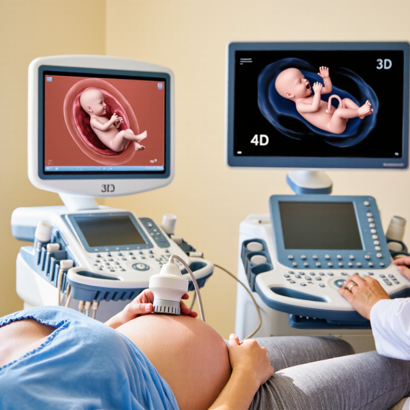
In pregnancy care, 3D ultrasound creates a still, three-dimensional image from many sound-wave angles. It can show facial contours, limbs, and certain surface details more clearly than a flat 2D image. A 4D ultrasound adds time, producing moving images of the fetus. You may see a hand open, lips move, or the baby turn.

The difference is mainly image presentation, not a separate level of fetal health assessment. Trained clinicians still rely on 2D ultrasound for growth, anatomy, fluid levels, and placental location. Medical guidance should determine the scan’s purpose. 3D imaging may support assessment of selected structural concerns, while 4D imaging can help observe movement in real time. Neither method replaces careful clinical interpretation.

Image quality depends on fetal position, gestational age, fluid volume, and the sonographer’s experience. A face pressed against the uterine wall may appear unclear. That can feel disappointing. It does not automatically suggest a problem. Non-medical scans should not delay recommended prenatal appointments or diagnostic testing. Ask what the examination can realistically answer, who will interpret it, and how the findings enter your maternity record. More detail is not always more useful. Sometimes a clear 2D view gives the clinician the most reliable information.

How 3D and 4D Ultrasound Technologies Work

During pregnancy, 3D and 4D ultrasound use the same basic principle: high-frequency sound waves create images inside the uterus. A transducer sends sound through a layer of gel. Returning echoes reveal boundaries between tissues, fluid, and the developing baby. The process uses no ionizing radiation.

3D Ultrasound 3D ultrasound collects many two-dimensional slices from different angles. Computer software combines them into a volume image. This can show facial contours, limbs, and surface details more clearly than a flat scan. However, the picture depends on fetal position, movement, fluid volume, and image quality. A hand covering the face can hide important details.

4D Ultrasound 4D ultrasound adds time to the 3D volume. It displays changing movement, such as a yawn, stretch, or turn, almost immediately. This makes the examination feel more vivid. It does not automatically provide better medical information. A trained sonographer still adjusts depth, angle, and gain carefully. The clinician may switch between 2D, 3D, and 4D views during one appointment.

It is easy to assume that the newest-looking image is the most useful. That assumption is not always correct. Medical decisions should rely on an appropriate examination and professional interpretation, not appearance alone. Families should ask what each view can realistically show at their stage of pregnancy. Sometimes the clearest answer remains a simple two-dimensional image.

Key Differences Between 3D and 4D Pregnancy Scans

How to Choose 4D vs 3D Ultrasound for Pregnancy 2026?

The key difference between 3D and 4D pregnancy scans is movement. A 3D scan creates a single, three-dimensional image of the baby. It may show facial features, hands, or the curve of the spine with greater depth than standard 2D imaging. A 4D scan adds time, producing moving 3D images, such as a yawn, stretch, or brief hand wave.

Neither option automatically provides a better medical assessment. Standard 2D ultrasound remains central for measuring growth, checking organs, and reviewing blood flow. Three-dimensional imaging can support specific evaluations, but its usefulness depends on the clinical question and the operator’s training. A baby facing the mother’s spine may produce unclear images, regardless of the selected scan.

Practical details matter. Amniotic fluid, fetal position, gestational age, and maternal tissue can affect image quality. Around the late second or early third trimester, facial images may look clearer, but timing varies. Ask a qualified ultrasound professional which method is clinically appropriate. Confirm who interprets the images and how results reach your maternity team. A beautiful moving picture can feel reassuring, yet appearance alone cannot confirm healthy development. The distinction is not always neat. Clear communication matters more than impressive footage.

When to Choose 3D or 4D Ultrasound During Pregnancy

Choosing 3D or 4D ultrasound during pregnancy depends on your clinical needs, timing, and expectations. A 3D scan creates still images of the baby’s surface, such as the face, hands, or feet. It may help families see clearer details when the baby is positioned well. A 4D scan adds movement, allowing you to watch actions like yawning, stretching, or opening the mouth in near real time.

For medical assessment, your qualified ultrasound professional should guide the decision. Standard 2D ultrasound remains central for checking growth, anatomy, fluid levels, and the placenta. A 3D scan may support evaluation of certain structural concerns, but it does not replace a complete medical examination. 4D imaging can be useful when movement matters, although it may not improve every diagnosis. The baby’s position, gestational age, and available imaging conditions all affect the result.

Timing also matters. Later in pregnancy, facial images may become harder to capture because space is limited. The baby may turn away, cover the face, or remain unusually still. It happens. A disappointing image does not automatically signal a problem. It may simply reflect timing or position. Ask what the scan is intended to assess, who will interpret it, and whether the service follows appropriate clinical safety procedures. A memorable picture is pleasant, but reliable medical information should remain the priority.

Safety, Cost, Availability, and Clinical Considerations

For a 2026 pregnancy scan, clinical purpose should guide the choice. Standard 2D ultrasound remains the primary diagnostic method. Three-dimensional imaging adds surface detail, while 4D displays that image in motion. The 2024 AIUM practice guidance states that ultrasound should use the lowest output and shortest exposure needed for a medical purpose. The FDA also reports a strong safety record for diagnostic ultrasound, while advising trained operators and prudent use. More images do not automatically mean better care.

Cost and availability differ sharply by location. Public maternity services may include medically indicated 2D scans, but elective 3D or 4D sessions often require self-payment. A 2024 market analysis from Grand View Research estimated the global 3D ultrasound market at over 3 billion dollars, reflecting expanding access, not equal access. Urban clinics may offer 4D appointments within days. Rural patients may face longer travel and fewer qualified sonographers. Prices can also exclude interpretation or follow-up imaging. Ask for the full fee.

Choose 3D or 4D when a clinician believes it could clarify anatomy or support a specific assessment. For routine reassurance, the extra detail may add little. Facial images can look impressive. They can also create false confidence. ACOG recommends medically indicated examinations performed by appropriately trained professionals, rather than souvenir scans. I would question any clinic promising certainty from a single picture. Availability, operator skill, gestational age, fetal position, and maternal body habitus all affect image quality.

Both 2D and 3D/4D ultrasound use non-ionizing sound waves and are considered safe when medically justified and performed by trained professionals. 2D ultrasound remains the standard for routine diagnostic assessment, while 3D/4D is generally an optional adjunct for selected anatomical or visualization needs. Relative cost and availability vary by location, but 3D/4D examinations are commonly less available and may cost more.

FAQS

How do 3D and 4D ultrasound scans create images?

A transducer sends high-frequency sound waves through gel. Returning echoes form images of tissues, fluid, and the developing baby. No ionizing radiation is used.

What makes 3D ultrasound different from standard 2D ultrasound?

3D ultrasound combines many two-dimensional slices into one volume image. It can show facial contours, hands, feet, and surface details more clearly.

What does 4D ultrasound add?

4D ultrasound adds movement to a 3D image. You may see yawning, stretching, mouth opening, or turning almost immediately.

Does 4D ultrasound always provide better medical information?

No. A moving image may feel more vivid, but it does not guarantee a better diagnosis. Sometimes, a simple 2D image answers the clinical question best.

When might 3D or 4D ultrasound be useful?

It may help assess certain structural concerns or show surface details. A qualified ultrasound professional should decide whether it is appropriate.

Can the baby’s position affect the scan?

Yes. A hand may cover the face. The baby may turn away or remain still. Position matters.

When is the best time for clear facial images?

Timing depends on gestational age, fluid volume, and fetal position. Later in pregnancy, limited space can make facial images harder to capture.

Does a poor image mean something is wrong?

Not necessarily. The baby may be facing away, moving, or covering the face. A disappointing picture is not automatically a medical concern.

What should families ask before choosing a 3D or 4D scan?

Ask what the scan will assess and who will interpret it. Also ask whether appropriate clinical safety procedures are followed.

Should families judge the scan by appearance alone?

No. The newest-looking image may not be the most useful. I might expect more from a vivid picture, but appearance cannot replace professional interpretation.

Conclusion

Choosing between 3D and 4D ultrasound during pregnancy depends on your medical needs, timing, budget, and local availability. 3D ultrasound creates detailed still images of the baby’s surface, while 4D ultrasound adds real-time movement, allowing parents and clinicians to observe facial expressions, body motion, and changing positions. Many people ask, “why is 4d ultrasound better than 3d for pregnancy,” but 4D is not automatically better. Its main advantage is motion, whereas 3D may provide sufficient detail for viewing selected anatomical features and can be more affordable or accessible.

Both technologies use sound waves and should be performed by trained professionals for appropriate medical reasons. Standard diagnostic ultrasound remains the priority for monitoring growth, development, and pregnancy health. The best choice may depend on the gestational stage, the position of the baby, image quality, examination purpose, cost, and healthcare provider recommendations. Expectant parents should discuss their goals and any concerns with a qualified maternity professional before scheduling an elective scan.

Oliver

Oliver

Oliver is a seasoned marketing professional with a wealth of expertise in driving brand awareness and engagement. With a deep understanding of our company's product offerings, he consistently delivers high-quality content that enriches our professional blog. His insights not only shed light on......